*This article by Dr. Stuart Foster was published as part of the old TFRI blog.

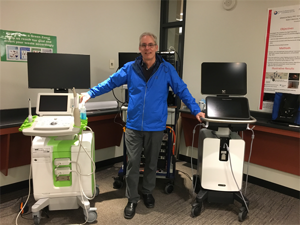
Dr. Stuart Foster poses with some of his team's groundbreaking prostate imaging technology.
Dr. Stuart Foster is a senior scientist at Toronto’s Sunnybrook Research Institute, as well as professor and Canada Research Chair in ultrasound imaging, Department of Medical Biophysics, University of Toronto. His team, which currently includes Drs. Christine Demore, Brian Wilson, and Gang Zheng (Princess Margaret Cancer Centre), specializes in innovative new imaging techniques, and has been funded by the Terry Fox Research Institute since the 1980s. In this blog post, Dr. Foster details the ground breaking work the group is doing in prostate cancer imaging through The Terry Fox New Frontiers Program Project Grant on Nano Particle Enhanced Photoacoustic Imaging for Cancer Localization and Therapeutic Guidance – and how the team hopes to improve treatment for patients.
“The prostate is probably one of the most difficult organs in the body to image, even with MRI or PET imaging, or ultrasound – it’s challenging, and that’s just the way it is. Traditional prostate imaging with ultrasound is generally done with a device called the transducer, which has an operating frequency in the range of eight megahertz or eight million oscillations per second. It’s been used for years and years, but has limitations. Our team has changed the game a bit with prostate imaging by increasing the frequencies we use from eight megahertz to about 20, which improves the resolution by a factor of 2.5 or three and gives you a much clearer picture of the prostate with ultrasound.
We’ve also capitalized on a technology called photoacoustic imaging (PAI), which is a combination of optical imaging and ultrasound imaging that gives you information unavailable by either technique individually. PAI works by emitting a very short burst of light in the order of 10 nanoseconds long into the tissue. When that light interacts with things that are absorbing light in the tissue, like red blood cells, those areas heat up instantaneously, and this heat causes expansion of the tissue. Then we use an ultrasound transducer to read out that information, so it’s a two-step process: excitation of the tissue with laser, and then readout with ultrasound. The reason this is a powerful approach is that it allows us to take advantage of the fantastic information that’s available through optical technologies – for example the absorption of light in tissue (spectroscopy) – and the ability of ultrasound to do readouts at significant depths. By combining these two things we in theory get the best of both worlds.
Technology developed with the support of the Terry Fox Research Institute and Foundation has been translated as a fully commercialized system that’s now been used in a multi centre clinical trial involving over 1000 patients. At the same time, we’re working on fundamental improvements of PAI at the basic level as part of our TFRI New Frontiers Program – and that really means “new frontiers” not research that has been done or exists or has been proven clinically. One exciting area of investigation is based on developments in Dr. Gang Zheng’s lab where he has discovered a type of biodegradable nano particle called a porphysome. Porphysomes are strong absorbers of infrared light so that they can act as either a photoacoustic beacon or, when illuminated with higher intensity light, a source of heat to ablate tumour tissue. The ability to image and treat with the same agent is often referred to as a theranostic: both a therapy and a diagnostic rolled into one. Porphysomes can also be modified to create a “targeted agent” that will seek out and bind important prostate markers such as prostate specific membrane antigen (PSMA). This may enable much more specific treatments.
Here’s how we envision this PAI theranostic technology might work. Let’s imagine we have this high-tech, super high frequency probe with optical fibres that deliver the short pulses of light energy inside the rectal cavity, and that light is projected towards the prostate. And then, at the same time, we could conceivably even introduce other optical fibres in the urethra to illuminate the prostate from its centre. By then injecting the targeted porphysome contrast nano particles into the blood stream you have an injectable tracer that will find a target in the prostate and you can read that information out with the photoacoustic system, and that would bring the whole concept of molecular imaging of the prostate with ultrasound into the realm of possibility. An example of a possible target is PSMA -- having found the target, it may then be possible to perform photothermal therapy by taking advantage of porphysomes’ inherent theranostic potential.
We’re really pushing the envelope on a variety of very interesting imaging techniques that might allow us to get a better picture of how prostate tumours are developing, and eventually how molecular aspects of prostate cancer change as either a therapy is introduced or as the cancer progresses or hopefully regresses. I hope this project will result in better treatment options for prostate cancer patients. One of the difficult questions these patients face is: do they get their prostate taken out and accept all of the nasty things that might also happen as a result of that operation, or do they try watchful waiting and see if anything happens, or do they use an imaging technology to see if a prostate tumour is aggressive or something that you can leave for a long period of time with no problems. With better imaging tools like PAI and better contrast agents like porphysomes we might be able to answer that question and save the health care system a whole lot of unnecessary operations - why take people’s prostates out if they don’t need to come out? Better information helps make better decisions.”